- This yields glucose for the muscles
- This yields fatty acids for the muscles
The PKA pathway
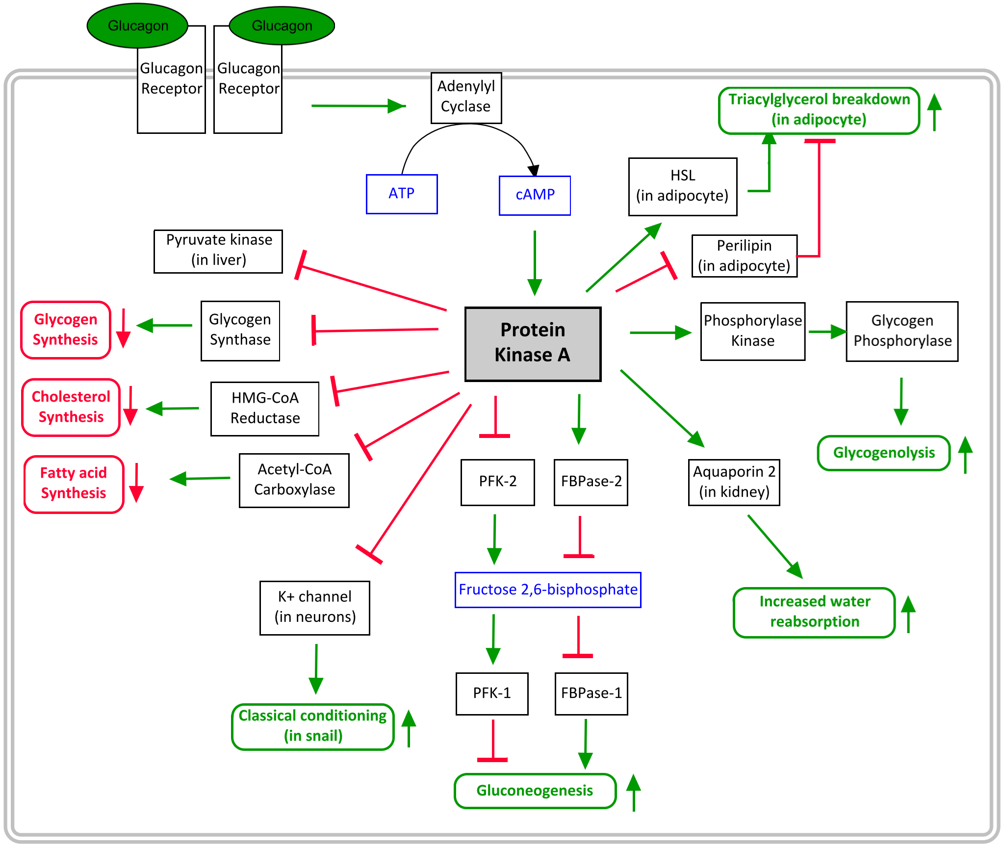
Both glucagon and epinephrine increase gluconeogenesis and glycogenolysis, and they inhibit the utilization of glucose by the liver. The two hormones achieve theses similar effects by acting through very similar pathways.
Differences between glucagon and epinephrine pathways
Glucagon acts only on the liver, while epinephrine acts on muscle, liver, as well as other tissues.
Notably, PKA inhibits L-type pyruvate kinase (which is in the liver) but does not influence pyruvate kinase in the muscle. This causes glucagon and epinephrine to inhibit glycolysis in the liver.
From receptor binding to PKA activation
Both glucagon receptor and the β2-adrenergic receptor are G-protein coupled receptors. When the hormone binds to them, the receptor binds activates the membrane-bound enzyme called adenlylyl cyclase. This enzyme catalyses the reaction of converting ATP to cAMP. When the level of cAMP in the cell increases, a protein called Protein Kinase A is activated. PKA is what mediates the cells response.
After PKA activation
PKA then phosphorylates many different enzymes. This either increases or decreases their activity, depending on the enzyme.
Remember that epinephrine and glucagon are hormones that signal for the body to raise the blood glucose level, while decreasing the amount of glucose the liver uses. With this is mind, it’s easier to remember what PKA affect. PKA activates enzyme related to gluconeogenesis, β-oxidation and glycogen breakdown, while inhibiting enzymes that are related to pathways that use energy for other things than gluconeogenesis, like cholesterol synthesis, fatty acid synthesis and glycogen synthesis.
After a while, the enzyme called cyclic nucleotide phosphodiesterase or simply PDE, will degrade cAMP to 5’-AMP, which will decrease the level of cAMP in the cell, which will decrease the effect of PKA, eventually reversing the effects of the hormone.

The PKA pathway and the proteins PKA affects. More details here.
Desensitization of the β-adrenergic receptor
The β-adrenergic receptor can be desensitized, which means that the cell becomes less sensitive to the hormone after being exposed to the hormone for a long time.
After the receptor has bound an epinephrine, a protein called βARK will phosphorylate the receptor, which inactivates it. Following this, another protein called β-arrestin, or βarr, will bind to the phosphorylated receptor, and remove the whole receptor from the cell surface by endocytosis. As the receptor isn’t on the surface anymore, but rather inside a vesicle inside the cell, it obviously can’t bind epinephrine anymore. The receptor is now desensitized.
After a period, βarr dissociates, the receptor is dephosphorylated, and the receptor is returned to the cell surface.
Summary
- What is the purpose of glucagon, and where is it synthesised?
- Glucagon upholds the blood glucose level in periods of fasting
- It’s synthesised by alpha cells in the Langerhans islets
- Glucagon is released during fasting
- Tyrosine -> L-DOPA -> Dopamine -> Norepinephrine -> Epinephrine
- Needs vitamin C, adoMet, THB and PLP.
- During periods of stress
- The protein kinase A pathway
- Glucagon binds to glucagon receptor or epinephrine binds to β-adrenergic receptor, both of which are G-protein coupled receptors
- This stimulates adenylyl cyclase, which synthesises cAMP
- cAMP activates PKA
- Glucagon acts only on the liver, while epinephrine acts on muscle, liver, as well as other tissues.
- Both inhibit L-type pyruvate kinase in the liver, thereby inhibiting glycolysis there
- Triacylglycerol breakdown
- Glycogenolysis
- Gluconeogenesis
- Glycogen synthesis
- Cholesterol synthesis
- Fatty acid synthesis
- βARK phosphorylates the receptor, and βarr removes it from the cell surface by endocytosis
